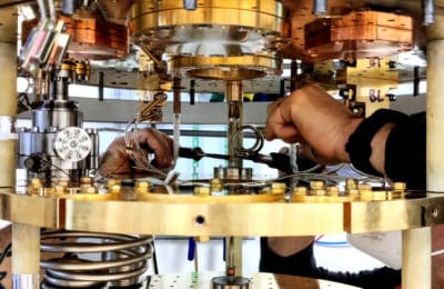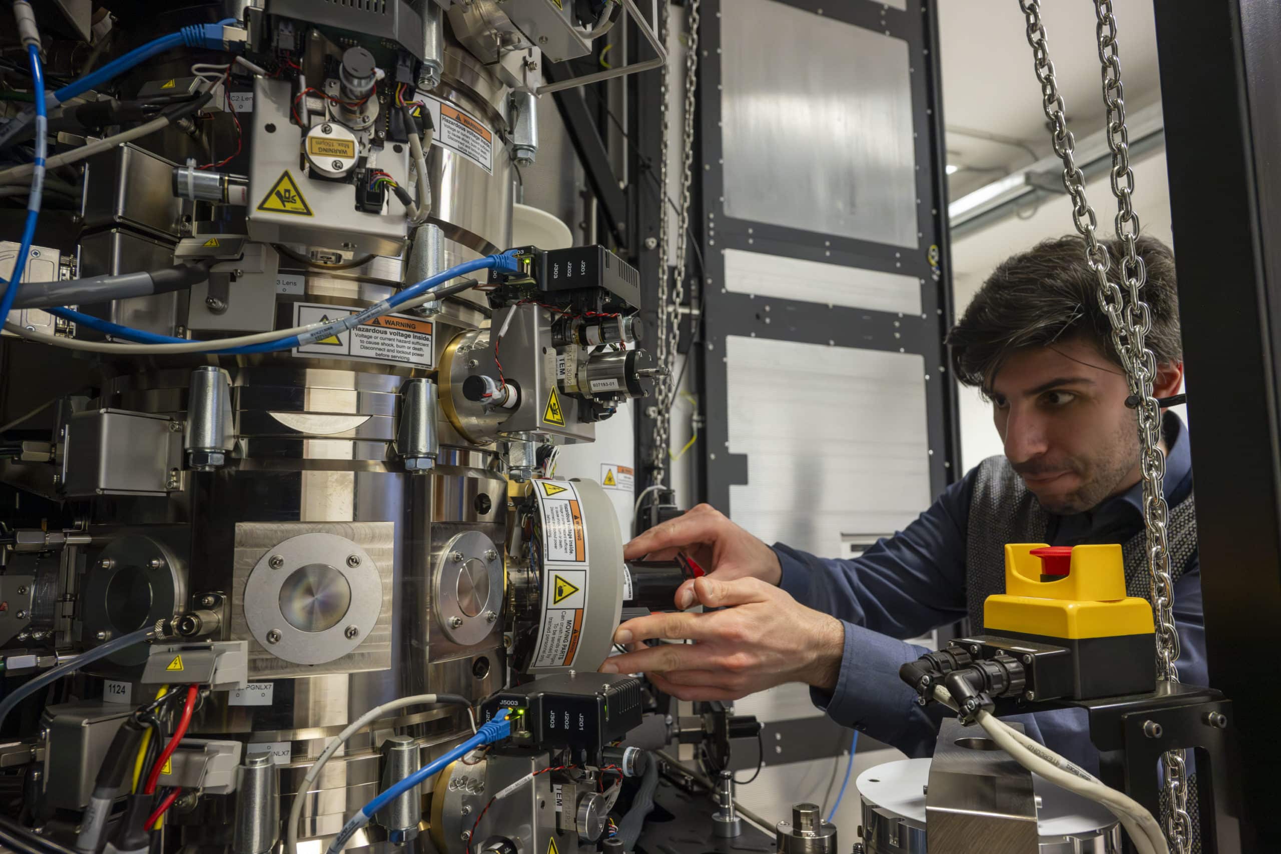
A research team has demonstrated, for the first time, how to detect the angular momentum of electrons in solids after they transferred energy to an atomic transition using an advanced transmission electron microscope. The study, published in Nature Communications and titled Demonstration of angular-momentum-resolved electron energy-loss spectroscopy, represents a significant step forward in the use of electron microscopy to probe electronic behavior in materials at the nanoscale.
The work was led by Vincenzo Grillo, with Giovanni Bertoni, Enzo Rotunno, Paolo Rosi and Gian Carlo Gazzadi (Cnr Nano), in collaboration with Stefano Frabboni (Unimore), and researchers from Cnr-ISMN, the Ernst Ruska-Centre for Microscopy and Spectroscopy with Electrons (Forschungszentrum Jülich), the University of Ottawa, and Thermo Fisher Scientific.
“This is the first time we can read the direction and symmetries of electron orbits inside a material,” says Vincenzo Grillo. “It’s a bit like measuring how a spinning top is spinning, but on the scale of atoms.”
Unlike conventional techniques that detect only the energy of inelastically scattered electrons, this method captures both energy and angular momentum. This dual detection approach enabled researchers to distinguish between different types of chemical bonds—even when they overlap in energy—providing a new tool for investigating quantum phenomena in complex materials. The method was validated on a thin crystal of boron nitride, and the results mark the first experimental detection of angular momentum in this context.
The experiment was designed and developed at Cnr Nano, including the fabrication of key optical components and the implementation of the computational routines required to analyze the data.
This result opens promising perspectives for efficient, atomically resolved studies of magnetic properties and chiral phenomena, such as magnetic chiral dichroism. Future work will focus on improving spatial resolution and applying this technique to magnetic and functional materials, with the goal of mapping electron behavior with atomic precision in complex, real-world systems.
Original paper: Tavabi, A.H., Rosi, P., Bertoni, G. et al. Demonstration of angular-momentum-resolved electron energy-loss spectroscopy. Nat Commun 16, 6601 (2025). https://doi.org/10.1038/s41467-025-60804-3